Shoulder Joint Anatomy Diagram : Joints - HUMAN ANATOMY WEB SITE : The shoulder joint is the connection between the chest and the upper extremity.. Dislocation of the shoulder is extremely painful and may require surgical repair or even cause permanent damage. Various types of injuries and degenerative conditions can cause the shoulder to become painful. 2.bones of shoulder joint 3.joints of the shoulder complex glenohumeral acromioclavicular sternoclavicular scapulothoracic 4.muscles of the shoulder. Like all other joints, the shoulder is first examined by obtaining a baseline study consisting of two views perpendicular to each radiographic anatomy. It can help you understand our world more detailed and specific.
Diagram of the different insertions of the anterior capsule as seen on the axial plane (arrowheads). The shoulder joint is vulnerable to dislocations from sudden jerks of the arm, especially in children before strong muscles have developed. 7 draw labelled diagram showing the relations of shoulder joint. Human anatomy diagrams and charts show internal organs, body systems, cells, conditions, sickness and symptoms information and/or tips to ensure one lives in good health. Kinesiology & anatomy chapter 10 shoulder joint.
Shoulder Joint X-Ray from www.edoctoronline.com The deepest layer of the shoulder includes the bones and the joints. Various types of injuries and degenerative conditions can cause the shoulder to become painful. The shoulder joint is the connection between the chest and the upper extremity. 6 describe briefly the abduction at shoulder joint. 8 name the arteries and the. The shoulder joint (glenohumeral joint) is a ball and socket joint between the scapula and the humerus. The chest, upper back and shoulder connect the upper limb to the trunk of the body and control it's movements.the clavicle connects to the sternum via the sternoclavicular joint and to the scapula by the acromioclavicular joint. Three bones come together at the shoulder joint.
8 name the arteries and the.
Assessment | biopsychology | comparative | cognitive | developmental | language | individual differences | personality | philosophy | social | methods | statistics | clinical | educational | industrial | professional items | world psychology |. The next layer is made up of the ligaments of the joint capsule. The anatomical basis of clinical practice. The shoulder joint is one of the most movable joints in the human body. We hope this picture right shoulder joint anterior and posterior view can help you study and research. For more anatomy content please follow anatomy is the amazing science. Superficial muscle that covers the shoulder joint on three sides, giving the shoulder its characteristic rounded shape. This diagram here just shows the joint capsule itself. Human anatomy diagrams and charts show internal organs, body systems, cells, conditions, sickness and symptoms information and/or tips to ensure one lives in good health. All four joints work collectively together to achieve normal shoulder girdle movements. Simple easy notes for quick revision for exams. Shoulder anatomy the shoulders are made up of bones cartilage muscles tendons and ligaments. Just remember the articulating surfaces.
Three bones come together at the shoulder joint. 2.bones of shoulder joint 3.joints of the shoulder complex glenohumeral acromioclavicular sternoclavicular scapulothoracic 4.muscles of the shoulder. Shoulder abduction is any motion of the shoulder joint that involves lifting your arm away from the body. Shoulder joint is the most mobile joint of the human body. Body of the scapula free of superimposed ribs.
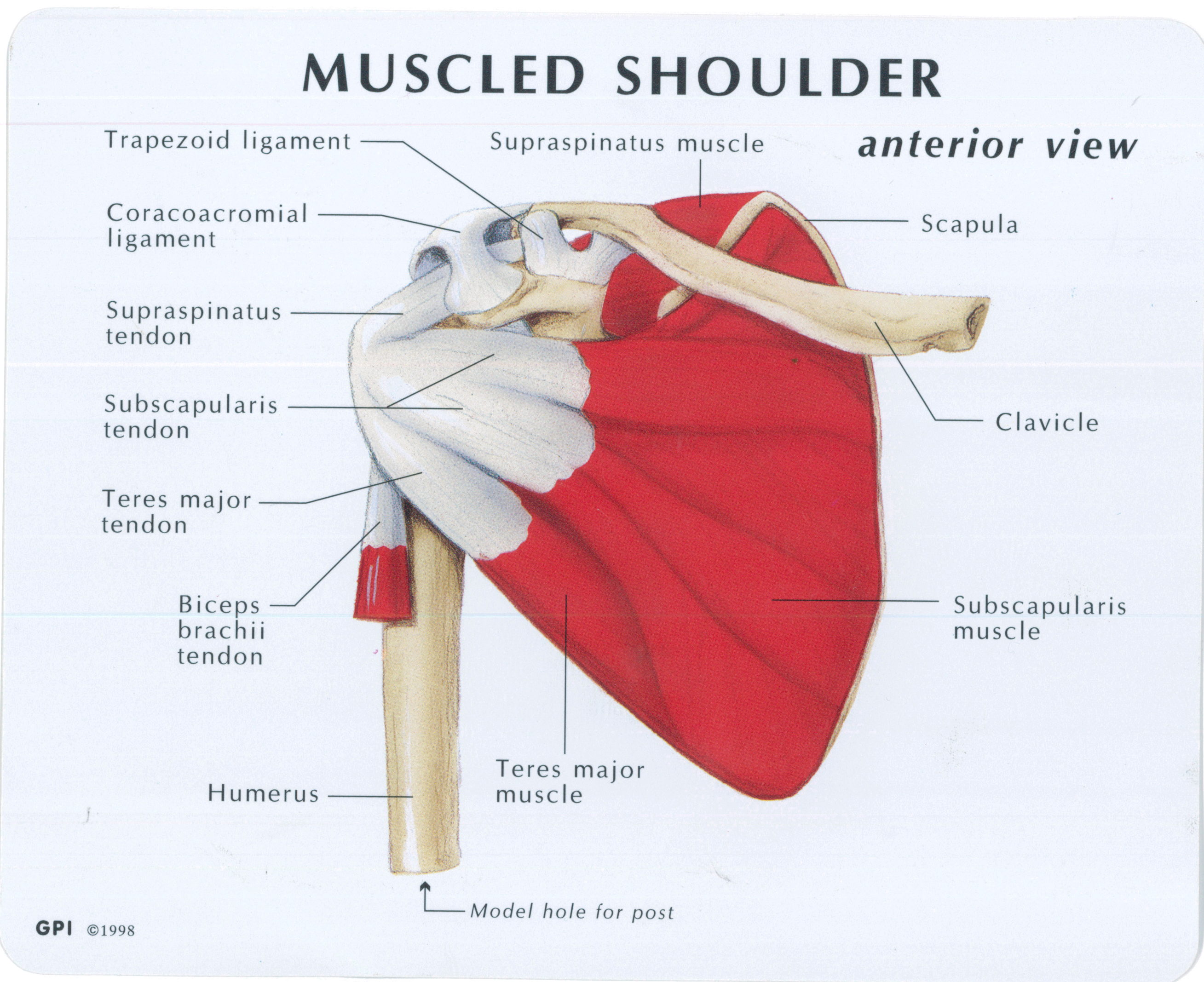
Muscled Shoulder Joint Model - MedWest Medical Supplies from www.medwest.ca The shoulder anatomy includes the anterior deltoid, lateral deltoid, posterior the rotator cuff is a complex and delicate structure of the shoulder anatomy. The posterior deltoid's purpose is to extend learning about the anatomy of your shoulders is key to preventing shoulder injuries. Diagram of the different insertions of the anterior capsule as seen on the axial plane (arrowheads). Kinesiology & anatomy chapter 10 shoulder joint. It is located between the shoulder and the elbow joint. Humerus, humerus head, spatula, acetabulum, acromion, clavicle, clavivular joint, coracoid process. It is the major joint connecting the upper limb to the trunk. It allows the upper limb to have a wide array of movements.
Shoulder abduction is any motion of the shoulder joint that involves lifting your arm away from the body.
It is the major joint connecting the upper limb to the trunk. The anatomical basis of clinical practice. 1.introduction shoulder joint is formed by scapula and clavicle (which is also called as shoulder girdle)and proximal humerus. The chest, upper back and shoulder connect the upper limb to the trunk of the body and control it's movements.the clavicle connects to the sternum via the sternoclavicular joint and to the scapula by the acromioclavicular joint. By learning how each part works, you can better understand how. Kinesiology & anatomy chapter 10 shoulder joint. Assessment | biopsychology | comparative | cognitive | developmental | language | individual differences | personality | philosophy | social | methods | statistics | clinical | educational | industrial | professional items | world psychology |. This human anatomy diagram with labels depicts and explains the details and or parts of the anatomy of the shoulder joint. It allows the upper limb to have a wide array of movements. Posted on december 13, 2018december 12, 2018. We hope you will use this picture in the study and. In common usage, shoulder joint mostly refers to the glenohumeral joint, the major joint of the shoulder but can also include acromioclavicular joint. Shoulder anatomy the shoulders are made up of bones cartilage muscles tendons and ligaments.
Shoulder injections injections to the shoulder can be performed either for diagnostic purposes or for aspiration of joint fluid. This human anatomy diagram with labels depicts and explains the details and or parts of the anatomy of the shoulder joint. Wiring diagram for genie garage door opener. In this article, we shall look at the anatomy of the shoulder joint and its important clinical correlations. In human anatomy, the shoulder joint comprises the part of the body where the humerus attaches to the scapula.1 there are two kinds of cartilage in the joint.
A Frozen Shoulder in a Hot Summer - by Raphaëlle Strub from www.healthybynaturecalgary.ca Shoulder joint of human body anatomy infographic diagram with all parts including bones ligaments muscles bursa cavity capsule cartilage membrane for medical science education and health care. Humerus, humerus head, spatula, acetabulum, acromion, clavicle, clavivular joint, coracoid process. 2.bones of shoulder joint 3.joints of the shoulder complex glenohumeral acromioclavicular sternoclavicular scapulothoracic 4.muscles of the shoulder. Knowing the basic anatomy and surface landmarks of the shoulder for the subacromial space, glenohumeral joint, and ac joint is critically important for safe and effective… Home > blog > anatomy > shoulder anatomy: The deepest layer of the shoulder includes the bones and the joints. The chest, upper back and shoulder connect the upper limb to the trunk of the body and control it's movements.the clavicle connects to the sternum via the sternoclavicular joint and to the scapula by the acromioclavicular joint. Assessment | biopsychology | comparative | cognitive | developmental | language | individual differences | personality | philosophy | social | methods | statistics | clinical | educational | industrial | professional items | world psychology |.
In this article, we shall look at the anatomy of the shoulder joint and its important clinical correlations.
In human anatomy, the shoulder joint comprises the part of the body where the humerus attaches to the scapula.1 there are two kinds of cartilage in the joint. This human anatomy diagram with labels depicts and explains the details and or parts of the anatomy of the shoulder joint. This incongruent bony anatomy allows for the wide range of movement available at the shoulder joint but is also the reason for the lack of joint stability. The first type is the white cartilage on the ends of the bones (called articular cartilage) which allows the bones to glide and move on each other. 2.bones of shoulder joint 3.joints of the shoulder complex glenohumeral acromioclavicular sternoclavicular scapulothoracic 4.muscles of the shoulder. Equally extensive are the muscles affecting the shoulder movement, including: In this article, we shall look at the anatomy of the shoulder joint and its important clinical correlations. It is located between the shoulder and the elbow joint. This mobility provides the upper extremity with tremendous range of motion such as adduction, abduction, flexion, extension, internal rotation, external rotation, and 360° circumduction in the shoulder joint anatomy. It allows the upper limb to have a wide array of movements. Comprising of numerous ligamentous and muscular structures, the only the joint capsule attaches proximal to the glenoid fossa and attaches further distally to the anatomical neck of the humerus. 7 draw labelled diagram showing the relations of shoulder joint. Diagram of the different insertions of the anterior capsule as seen on the axial plane (arrowheads).
Wiring diagram for genie garage door opener shoulder anatomy diagram. Click now and learn everything about its anatomy and function at kenhub!


Posting Komentar
0 Komentar